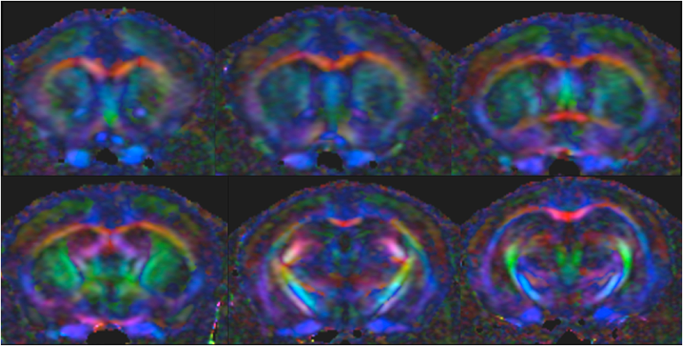
Diffusion-weighted imaging (DWI) MRI is indispensable for the study of white matter structures in the brain. Many DWI studies of the mouse brain have been performed using ex vivo imaging which provides high spatial resolution images but requires long acquisition times which are not suitable for in vivo imaging.
CAI researchers have now developed an optimised protocol for DWI of mouse brain in vivo at ultra-high magnetic field (16.4T) that combines high in-plane spatial resolution with acceptable acquisition times (Alomair et al., PLoS ONE, 2015). This protocol will be useful for longitudinal study of animal models of neurological disease, in particular those suffering from white matter changes.
PLoS ONE, 2015 June 25. doi: 10.1371/journal.pone.0130133.