Ultrasound
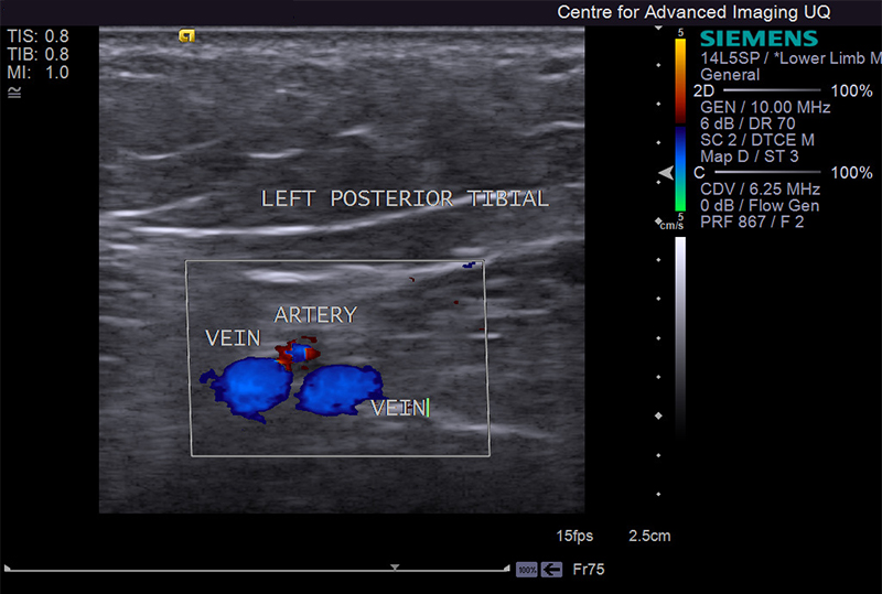
The Centre is equipped with a high performance Siemens/Acuson S3000 ultrasound scanner. The ultrasound can be used for a wide variety of studies including long structures such as tendons, nerves and blood vessels, fetal monitoring, the assessment of musculoskeletal structures, heart valve function, the excursion of a tendon or the contraction of a muscle, blood flow velocity and direction, as well as organ perfusion.
This latest generation system has the following features:
- B-mode, colour and pulsed wave Doppler, power Doppler, harmonic imaging
- Spatial compounding in B mode, colour and power Doppler
- Capability of multimodal review – for example to compare the ultrasound image to that of MRI/CT/PET on the same screen.
- Panoramic imaging of 240cm in length
- Off line transfer and data processing
- Elastography capabilities to assess tissue stiffness. This system is capable of real time voxel placement and shear wave velocity estimation, as well as the generation of corresponding colour coded tissue stiffness map.
For any enquiries about this equipment, please contact Gail Durbridge.