Animal Imaging
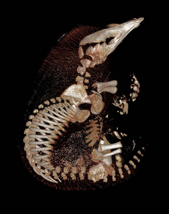
The Centre for Advanced Imaging's pre-clinical animal systems provide access to image analysis for cells, tissues, small animals and multi-modal imaging. Our comprehensive range of imaging technologies and expertise enable novel small and large animal research.
The primary capabilities of CAI's animal facilities are structure, function and molecular imaging in live animal, fixed tissue or materials. The imaging equipment is contained in a PC2 facility with animal holding facilities, anaesthetics, and tissue and sample preparation facilities.
Magnetic Resonance Imaging (MRI) has become an essential research tool as it provides detailed image acquisition from soft tissue in situ, without sample destruction or ionising radiation. Detailed MRI brain images are universally recognised, however this is only a fraction of the potential of MRI.
Beyond structural detail, MRI can provide:
- measurement of flow and perfusion of tissue, including changes in vascularity, an essential tool for determining tumour growth and a potential biological marker for assessing the efficacy of tumour treatments.
- cellular density, organisation and connectivity. Diffusion imaging can used to develop ‘roadmaps’ of connection pathways in the brain and is also useful to study muscle tissues and cancer diagnosis in prostate and breast tissues.
- non-destructive measurement of metabolites in vivo using magnetic resonance spectroscopy, allowing longitudinal studies of metabolic processes.
- ATP, PCr, Pi and intracellular pH, essential for regulation of cellular energetics, can be probed using phosphorus MR.
- salt balance in tissue, in particular the intracellular/extracellular sodium balance, using sodium MR.
- the metabolism of 13C labelled biochemicals can be followed using carbon MR.
For more information about using the CAI animal imaging facilities in your research download the facility brochure (below) or contact the Facility Manager.