Anomalous Relaxation and Diffusion Study Group
Live meetings fortnightly
The Anomalous Relaxation and Diffusion Study Group aims to bring together people interested in the use of traditional and anomalous analytical methods linking with biologically influenced changes in tissue. For example, the group uses anomalous relaxation and diffusion models and apply these in NMR/MRI for the purpose of extracting additional information on tissue microstructure. Whilst we have a keen interest in extracting information from biomedical imaging data, it does not preclude other areas where analytical methods play an important role in extracting information from data collected.
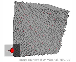 If you have an interest in using mathematical models, or artificial intelligence or machine learning approaches, for probing tissue microstructure and constituents, or just in using models for extracting novel information from biologically relevant data, then you may wish to join the Study Group to keep up to date with recent activities within this area.
The Study Group meets fortnightly via Zoom. The live session involves a 30-40 minutes presentation by a speaker on a scientific topic chosen by them, followed by a 20-30 minutes discussion. Please follow the link to subscribe to the mailing list for notifications and registration details for upcoming webinars. Please note your details will not be used for any other purpose or shared with any other organisation.
We welcome contributions in the form of educational materials and research findings. Please contact Viktor Vegh if you would like to be added to the contributors list. All contributions will be made available via our website.
The webinar series will be back from September 2021 with another set of talks for the year. Please let a member of the Organising Commitee know if you would like to give a study group talk later this year.
View previously recorded webinars
Organising Committee
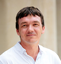 A/Prof Viktor Vegh, The University of Queensland, Australia
Viktor started off in the field of Electrical Engineering, before realising that his wholesome passion lied in mathematics. As such, he switched to a degree in Applied Mathematics at the Queensland University of Technology and completed with Honours. The experience was so positive that soon after completing this degree he enrolled in a PhD in Computational and Applied Mathematics at the same University. During this time, he equipped himself with skills such as advanced computational mathematics techniques, i.e. solution of large linear systems, electromagnetics and simulation of electromagnetic waves. These competencies allowed him to pursue a post-PhD post-doctoral fellowship at the Centre for Magnetic Resonance, the University of Queensland. His synergistic background in Electrical Engineering and Mathematics underpinned his initial contributions on instrumentation design and development in NMR/MRI.
It seems that people equipped with strong analytical skills seek out other opportunities during their career. Like Matt, Viktor pursued opportunities in finance and, decided to complete a Master of Commerce degree with a major in Applied Finance. Interestingly, his roots in mathematics led to the choosing of final year electives such as stochastic modelling, Brownian motion in relation to stock price movements, Monte Carlo methods, martingales and more computational mathematics with primary focus on finance models (e.g. Black-Scholes equation).
More recently, Viktor has focused on research and development of medical imaging methods for the direct in vivo mapping of biological effects, translatable to the diagnosis and monitoring of neurological diseases and disorders. As such, his research now focuses on understanding the underlying biological and physical processes influencing signal formation, specifically in MRI, via the use of mathematical models. Whilst his research focus has shifted over the years, it has always been, and continues to be, reinforced by his interests in the application of mathematics to real world problems. Applied finance remains a personal hobby for Viktor, in addition to cracking jokes with a straight face, offshore fishing and native Australian plants.
P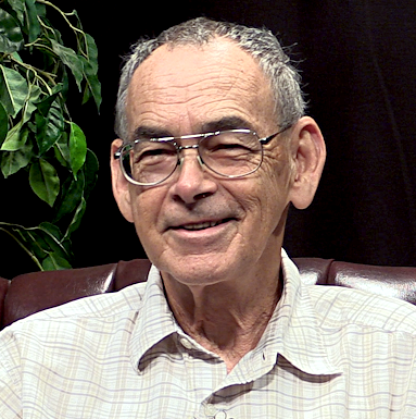 rof Richard Magin, University of Chicago Illinois, USA
Professor Magin was trained as an engineer (Ga. Tech), retrained in biophysics (Rochester), and then educated in the deep waters of biomedicine at the National Cancer Institute (NIH). He found work in the flat lands of East Central Illinois (UIUC) in bioacoustics and bio-electromagnetics. Tried to teach what he had learned – gaining knowledge that informed his dive into the cresting wave of MRI. Swimming there, he was surprised to find that most of what he knew a little about (relaxation and diffusion) could be fashioned into a boat for exploring and extracting imaging information suitable for the detection of cancer.
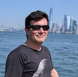 Dr Matt Hall, National Physical Laboratory, UK
Matt used to describe his career as Brownian, but now feels that anomalous might be a better description. He has a degree in Physics from the University of Sheffield, UK and a PhD in Mathematics from Imperial College London where he studied statistical mechanics applied to evolutionary ecology. He then spent two years working as a consultant in a City of London firm specialising in complexity science for business, where he developed logistics software for firms including the Danish postal service. He then discovered diffusion MRI, moving first to St George’s, University of London, and then UCL.
He spent some time developing Monte-Carlo simulation for diffusion MRI, and then went back to the City for a while, working at the Financial Services Authority – an experience which led him straight back to research. He moved back to UCL, firstly to CMIC and then to the UCL Great Ormond Street Institute of Child Health, where he worked on fractional approaches for diffusion in muscle tissue and Duchenne Muscular Dystrophy. His current incarnation is as a Principle Research Scientist at the UK’s National Physical Laboratory where he is developing MRI harmonisation and standardisation, including for clinical applications of fractional diffusion, advanced simulation-based methods and phantoms development.
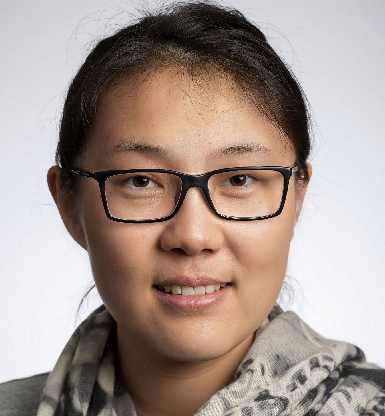 Dr Qianqian Yang, Queensland University of Technology, Australia
Qianqian is a senior research fellow in the School of Mathematical Sciences at Queensland University of Technology. She has extensive experience in developing computationally efficient methods for solving fractional order partial differential equations. With this background, her recent research interest lies in the application of these fractional-order models to real-world problems, especially in the area of using anomalous diffusion MRI models to probe brain tissue microstructure.
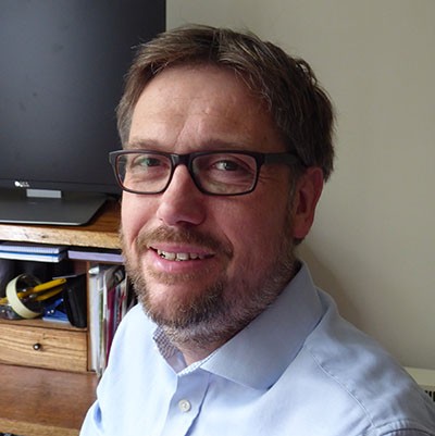 Prof Thomas Barrick (St George University London)
Tom has a degree in Mathematics from the University of Liverpool, UK (first class honours) and enjoyed the course sufficiently to do a Masters degree in Pure Mathematics (distinction) at the same university. After his Masters degree, while not knowing which direction to take his mathematical knowledge, he began a PhD in Medical Imaging at the Magnetic Resonance Image Analysis Research Centre (MARIARC), again at the University of Liverpool. This enabled him to begin his career in development of MRI techniques for image analysis and brain mapping.
He then moved to London to work on diffusion MRI and its application to patient diagnosis and disease progression at the Clinical Neuroscience Department at St George’s, University of London, UK. Eventually, after working with Matt Hall and meeting Richard Magin his interest in mathematics was rekindled in the form of fractional diffusion models and their applications in MRI. He currently works as an Associate Professor at St George’s, University of London and is developing Quasi-Diffusion Imaging, an advanced diffusion MRI technique based on fractional diffusion models that can be rapidly acquired in clinically feasible time.
 Dr David Reiter (Emory University)
David began his training in the field of mechanical engineering at North Carolina State University (USA). He completed a M.S. and PhD at the University of California at Davis as part of the Biomedical Research Group where his work focused on non-destructive measurements of mechanical strain in soft tissue using MRI. Inspired in graduate school by the versatility of MRI as a quantitative tool, he then completed a post-doctoral fellowship at the National Institute on Aging (NIA) in the National Institutes of Health (NIH), focusing on quantitative MRI and MRS towards improved non-invasive cartilage matrix characterization. He soon transitioned into clinical MRI research as a staff scientist and head of the clinical MRI facility at NIA. His work there supported aging studies using imaging and spectroscopy of multiple organ systems while his independent research examined modeling and analysis of multicompartment and anomalous relaxation and diffusion in musculoskeletal tissues like cartilage and muscle.
Now at Emory University in the Department of Radiology and Imaging Sciences and the Department of Orthopedics, his lab continues to develop quantitative MRI and MRS approaches for addressing basic and applied research questions primarily in the musculoskeletal system. These approaches aim to improve the measurement of biomechanical, metabolic, and functional tissue properties in healthy and diseased conditions with the goal of obtaining detailed characterization of tissue status and disease progression, and enhanced diagnostic accuracy.
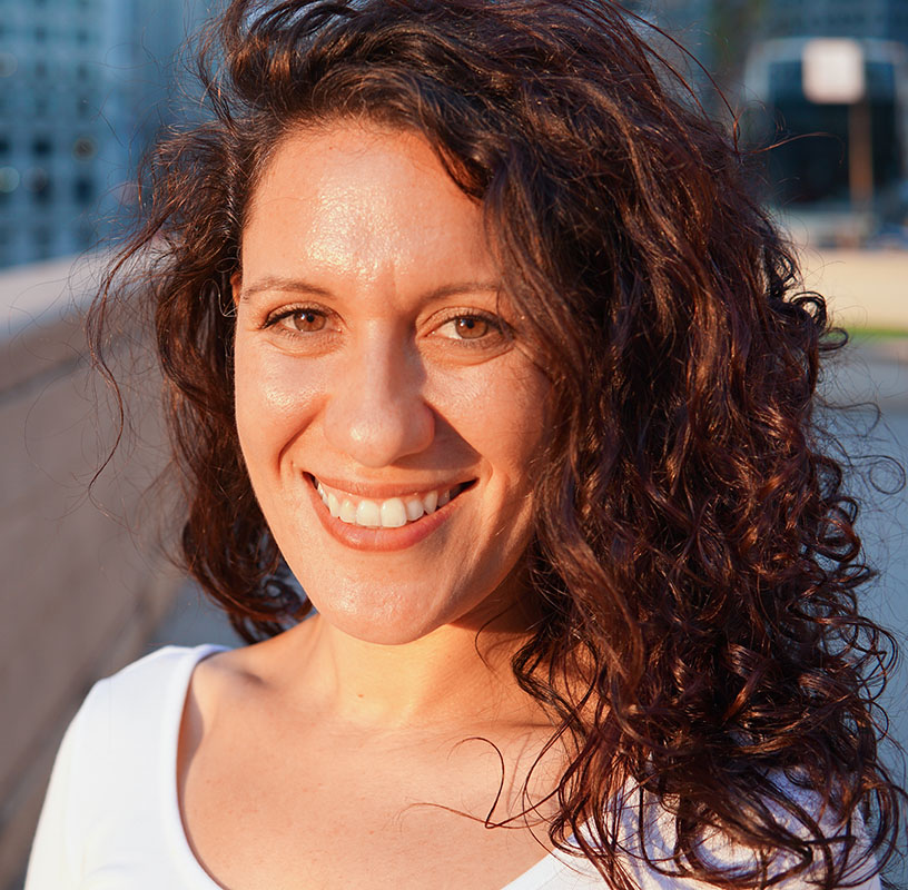 Dr Muge Karaman (University of Illinois, Chicago)
Muge received her first degree in Applied Mathematics from Istanbul Technical University in Istanbul (yes, that’s where Istanbul Technical University is located), Turkey (yes, that’s where Istanbul is located). She then pursued a Master’s degree in Computational Sciences and Engineering, where she worked on developing computational optimization models for disaster preparedness and response logistics; hoping to alleviate the impact of the expected powerful earthquakes in the region.
She found herself in the MRI field in 2009. She received her Ph.D. in Computational Sciences from Marquette University in Milwaukee, Wisconsin, where she had a lot of time to learn a lot about functional MRI (fMRI). Her research focused on developing complex-valued statistical models for fMRI and functional connectivity MRI in contrast to conventional techniques that use magnitude-only MRI data. As a reflection of her background in unconventional methods and models, she developed interest in anomalous diffusion after she joined the University of Illinois at Chicago (UIC) in 2014.
She is now a Research Assistant Professor at the Department of Bioengineering and the Center for MR Research of UIC. The overall goal of her research is developing and validating quantitative diffusion-weighted MRI techniques for the characterization of complex biological tissue in vivo, and integrating these techniques into computational models to address current and future medical challenges. She is particularly interested in investigating their feasibility in probing the underlying tissue characteristics in different disease conditions and organs, including adult and pediatric brain tumors, gastric cancer, and breast cancer. She is confident these advanced MRI techniques will facilitate critical clinical decisions in cancer diagnosis, early prediction of response to therapy, monitoring disease recurrence; and ultimately will help save lives.
Recent Publications
J. Gao, M. Jiang, R.L. Magin, R.G. Gatto, G. Morfini, A.C. Larson, and W. Li, Multicomponent diffusion analysis reveals microstructural alternations in spinal cord of a mouse model of amyotrophic lateral sclerosis ex vivo, PLoS ONE, 2020 (15): e0231598.
R.G. Gatto, C. Weissmann, M. Amin, A. Finkielsztein, R. Sumagin, T.H. Mareci, O.D. Uchitel, and R.L. Magin, Assessing neuraxial microstructural changes in a transgenic mouse model of early stage amyotrophic lateral sclerosis by ultra-high field MRI and diffusion tensor metrics, Animal Models and Experimental Medicine, vol. 3, pp. 117-129, 2020.
R.L. Magin, M.G. Hall, M.M. Karaman and V. Vegh, Fractional calculus models of magnetic resonance phenomena: relaxation and diffusion, Critical Reviews in Biomedical Engineering, vol. 48, pp. 285-326, 2020.
Q Yang, S Puttick, ZC Bruce, BW Day, V Vegh. Investigation of changes in anomalous diffusion parameters in a mouse model of brain tumour. Proceedings of Computational Diffusion MRI: International MICCAI Workshop 2019.
Q Yu, D Reutens, V Vegh. Can anomalous diffusion models in magnetic resonance imaging be used to characterise white matter tissue microstructure? NeuroImage 175: 122-137; 2018.
Qin, Shanlin, Liu, Fawang, Turner, Ian W., Yu, Qiang, Yang, Qianqian and Vegh, Viktor Characterization of anomalous relaxation using the time-fractional Bloch equation and multiple echo T2*-weighted magnetic resonance imaging at 7 T. Magnetic Resonance in Medicine, 77 4: 1485-1494 2016.
Yu, Qiang, Reutens, David, O'Brien, Kieran and Vegh, Viktor Tissue microstructure features derived from anomalous diffusion measurements in magnetic resonance imaging. Human Brain Mapping, 38 2: 1068-1081. doi:10.1002/hbm.23441 2017.
Y. Liang, S. Wang, W. Chen, Z. Zhou and R.L. Magin, "A survey of models of ultraslow diffusion in heterogeneous materials," Applied Mechanics Reviews, vol. 71, pp. 040802 (16), 2019.
R.L. Magin, M.M. Karaman, M.G. Hall, W. Zhu and X.J. Zhou, “Capturing complexity of the diffusion-weighted MR signal decay,” Magnetic Resonance Imaging, vol. 56, pp. 110-119, 2019.
R.L. Magin, H. Karani, S. Wang and Y. Liang, " Fractional order complexity model of the diffusion signal decay in MRI," Mathematics, vol. 7, pp. 348-364, 2019.
R.G. Gatto, A.Q. Ye, L. Colon-Perez, T.H. Mareci, A. Lysakowski, S.D. Price, S.T. Brady, M. Karaman, G. Morfini, and R.L. Magin, “Detection of axonal degeneration in a mouse model of Huntington’s disease: Comparison between diffusion tensor imaging and anomalous diffusion metrics,” Magnetic Resonance Materials in Physics, Biology and Medicine,” vol. 32(4), 461-471, 2019.
R.G. Gatto, M. Y. Amin, A. Finkielsztein, C. Weissman, T. Barrett, C. Lamoutte, O. Uchitel, R. Sumagin, T.H. Mareci and R.L. Magin, “Unveiling early cortical and subcortical neuronal degeneration in ALS mice by ultra-high field diffusion MRI," Amyotrophic Lateral Sclerosis and Frontal-temporal Degeneration, vol. 20, pp. 549-561, 2019.
TR Barrick, CA Spilling, C Ingo, J Madigan, JD Isaacs, P Rich, TL Jones, RL Magin, MG Hall, FA Howe. Quasi-diffusion magnetic resonance imaging (QDI): A fast, high b-value diffusion imaging technique. Neuroimage; 2020. May 1;211:116606. doi: 10.1016/j.neuroimage.2020.116606. Epub 2020 Feb 4.
C Ingo, TR Barrick, AG Webb, I Ronen (2017). Accurate Padé Global Approximations for the Mittag-Leffler Function, Its Inverse, and Its Partial Derivatives to Efficiently Compute Convergent Power Series. International Journal of Applied and Computational Mathematics 3: 347–362; 2017.
Reiter D.A., Magin R.L., Li W., Velasco M.P., Trujillo J.J., Spencer R.G. Anomalous T2 Relaxation in Normal and Degraded Cartilage. Magnetic Resonance in Medicine, September 2016, 76(3):953-962.
Selected as the “Editor’s Pick” for the issue; interview and video slides located here: http://www.ismrm.org/qa-with-david-reiter-and-richard-spencer/
Magin R.L., Li W., Velasco M.P., Trujillo J.J., Reiter D.A., Morgenstern A., Spencer R.G. Anomalous NMR relaxation in cartilage matrix components and native cartilage: Fractional-order models. Journal of Magnetic Resonance, June 2011, 210(2): 184-191.
Cameron, D., Reiter D.A.*, Bouhrara M.*, Fishbein K., Choi S., Bergeron C.M., Ferrucci L.*, Spencer R.G.* The Effect of Lipid Signal on Determination of Gaussian and Non-Gaussian Diffusion Parameters in Skeletal Muscle. NMR in Biomedicine, July 2017, 30(7):e3718. * signifies equal contributions.
Adelnia F., Cameron D., Bergeron C., Fishbein K.W., Spencer R.G., Reiter D.A., Ferrucci L. The Role of Muscle Perfusion in the Age-Associated Decline of Mitochondrial Function with Aging in Healthy Individuals. Frontiers in Physiology, 2019, 10:427
Reiter D.A., Spencer R.G., Xia Y. “Multi-component NMR Relaxation in Cartilage,” New Developments in NMR: Biophysics and Biochemistry of Cartilage by NMR and MRI, ed. Xia Y., and Momot K., Royal Society of Chemistry Publishing 2016.
Karaman MM, Sui Y, Wang H, Magin RL, Li YH, Zhou XJ: Differentiating Low- and High-grade Pediatric Brain Tumors Using a Continuous-time Random-walk Diffusion Model at High b-values. Magn. Reson. Med., 76:1149-1157, 2016.
Sui Y, Wang H, Liu G, Damen FW, Wanamaker C, Li Y, Zhou XJ. Differentiation of Low- and High-Grade Pediatric Brain Tumors with High b-Value Diffusion-weighted MR Imaging and a Fractional Order Calculus Model. Radiology 2015. 277(2):489-96.
Karaman MM, Wang H, Sui Y, Engelhard HH, Li YH, Zhou XJ: Fractional Motion Diffusion Model for Grading Pediatric Brain Tumors. NeuroImage: Clinical, 12:707-714, 2016.
Sui Y, Xiong Y, Xie KL, Karaman MM, Zhu W, Zhou XJ: Differentiation of Low- and High-Grade Gliomas Using High b-Value Diffusion Imaging with a non-Gaussian Diffusion Model. Am J Neuroradiol, 37(9):1643-1649,2016.
Tang L, Sui Y, Zhong Z, Damen FC, Li J, Shen L, Sun Y, Zhou XJ. Non-Gaussian diffusion imaging with a fractional order calculus model to predict response of gastrointestinal stromal tumor to second-line sunitinib therapy. Magn Reson Med. 2018 Mar;79(3):1399-1406.
Karaman MM, Zhou XJ. A fractional motion diffusion model for a twice-refocused spin-echo pulse sequence. NMR Biomed. 2018 Nov;31(11): e3960.
Zhang J, Weaver TE, Zhong Z, Nisi RA, Martin KR, Steffen AD, Karaman MM, Zhou XJ. White matter structural differences in OSA patients experiencing residual daytime sleepiness with high CPAP use: a non-Gaussian diffusion MRI study. Sleep Med. 2019 Jan;53:51-59. 20.
Zhong Z, Merkitch D, Karaman MM, Zhang J, Sui Y, Goldman JG, Zhou XJ. High spatial resolution diffusion MRI of Parkinson disease: lateral asymmetry of the substantia nigra. Radiology. 2019 April; 291(1):149-157.
Zhong Z, Merkitch D, Karaman MM, Zhang J, Goldman JG, Zhou XJ. Response: Is High-Spatial-Resolution Diffusion MRI Applicable for the Diagnosis of Parkinson Disease?. Radiology. 2019 May; (online).
Resources
Frontiers Research Topic
The overall goal of this Research Topic is to collate current MRI technology research and development and identify future imaging biomarkers of degeneration with potential for clinical translation. Both animal and human studies are welcomed, and where possible, findings should be supported by multi-faceted validation.
Special edition in Mathematics
The purpose of this Special Issue is to gather articles reflecting the latest developments of fractional calculus in the fields of nuclear magnetic resonance (NMR), electron spin resonance (ESR), and magnetic resonance imaging (MRI). Applications employing fractional calculus in the sub-disciplines of NMR/ESR spectroscopy, relaxation, diffusion, and MRI are encouraged.
|
Special Issue "Fractional Calculus in Magnetic Resonance" Dear Colleagues, The applications of fractional calculus in the field of magnetic resonance are widespread and growing. In particular, we can extend the capabilities of nuclear magnetic resonance (NMR), electron spin resonance (ESR), and magnetic resonance imaging (MRI) by the generalization of the integer-order derivatives found in the governing equations (Bloch, and Bloch–Torrey equations). |
Fractional Calculus Resources
(See for a list of books, journals and web sites)
|
Igor Podlubny :: Home > Fractional calculus > Resources Fractals and Fractional Calculus in Continuum Mechanics. CISM International Centre for Mechanical Sciences Series, vol. 378. Springer-Verlag New York, Inc., New York, 1997, 348pp, ISBN 321182913X. Chapter on Fractional Calculus by R. Gorenflo and F. Mainardi ; Chapter on numerical methods of Fractional Calculus by R. Gorenflo people.tuke.sk |
|
Fractional Calculus and Applied Analysis | De Gruyter Objective Fractional Calculus and Applied Analysis (FCAA, abbreviated in the World databases as Fract. Calc. Appl. Anal. or FRACT CALC APPL ANAL) is a specialized international journal for theory and applications of an important branch of Mathematical Analysis (Calculus) where differentiations and integrations can be of arbitrary non-integer order. |
|
Igor Podlubny :: Home > Fractional calculus > What is it? Fractional calculus: What is it? Fractional calculus is a generalization of ordinary differentiation and integration to arbitrary (non-integer) order. people.tuke.sk |
|
Welcome to www.fracalmo.org: this WEB site is developed by a research group interested in modelling processes in applied sciences (physics, engineering, finance, biology, ...) via mathematical methods based on fractional calculus.The name fracalmo originates from fractional calculus modelling.. Fractional calculus, in allowing integrals and derivates of any positive real order (the term ... |
Software resources
|
Igor Podlubny :: Home > Software Software: brief overview. FRACULUS toolbox: Fractional calculus toolbox for Matlab and Simulink – under development. Here are some parts that can be used separately: people.tuke.sk |
|
Kilbas and Saigo Function - File Exchange - MATLAB Central The Kilbas-Saigo model is defined by Equations (5) and (8) in the paper, "Fractional Models of Anomalous Relaxation Based on the Kilbas and Saigo Function," by E. Capelas de Oliveira, F. Mainardi, and J. Vaz Jr. and published in the journal, Meccanica 2014, Volume 49, Issue 9, pp 2049-2060. |
|
The Mittag-Leffler function - File Exchange - MATLAB Central Evaluation of the Mittag-Leffler function with 1, 2 or 3 parameters. Version 1.3.0.0 (11.7 KB) by Roberto Garrappa au.mathworks.com |
Education resources
The following lecture notes and MATLAB codes have been provided by Dr Qianqian Yang from the Queensland University of Technology and were originally delivered as part of the course “Numerical methods for solving space fractional reaction-diffusion equations” at the AMSI Winter School in 2019.
All the notes were written in MATLAB’s live scripts. Interested readers can change the parameters of a simulation and see the results in the same file. Reproduction, reuse, and distribution of these notes is not permitted without permission.
From random walks to fractional diffusion equations - Dr Qianqian Yang (Queensland University of Technology) AMSI Winter School 2019
Lecture Notes (.pdf)
Lecture Notes and Matlab codes (.zip)
Numerical methods for fractional diffusion-reaction equations - Dr Qianqian Yang (Queensland University of Technology) AMSI Winter School 2019
Lecture Notes (.pdf)
Notes and Matlab codes (.zip)
The Preconditioned Lanczos Method for Solving Fractional Diffusion-Reaction Equations - Dr Qianqian Yang (Queensland University of Technology) AMSI Winter School 2019
Lecture Notes (.pdf)
Notes and Matlab codes (.zip)
