16.4T MR imaging
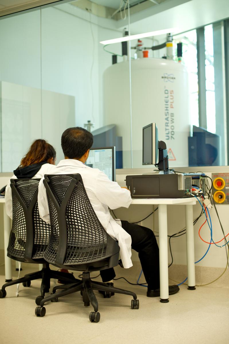
The University of Queensland commissioned a state-of-the-art 16.4T MRI spectrometer in October 2006. This powerful system is the only one of its kind in the southern hemisphere and one of six in the world.
The 700 MHz wide-bore micro-imaging system is capable of providing extremely detailed images of intact biological specimens. This spectrometer allows live mouse, fixed tissue and sample imaging from 4 mm to 30 mm diameter. Probes are available for imaging proton, fluorine and carbon.
Additionally, the Bruker Ultrashield Plus 700 WB Avance NMR spectrometer has a vertical open bore diameter of 89 mm. It has an available space of up to 34 mm diameter, which is optimal for live mouse imaging and various types of ex vivo samples.
The scanner is equipped with three types of gradient inserts: micro5, micro2.5 and mini0.5 which provide gradient strengths of 4.8, 2.5 and 0.5 mT/m/A.
To achieve highest sensitivity, the scanner is equipped with new generation SAW and quadrature microimaging coils (5, 10, 15, 20, 25, 28 and 35 mm diameters). The 5-15 mm coils are suitable for imaging of excised (fixed tissue samples) such as brain, spinal cord and biopsy samples. The 20-35 mm coils are dedicated for mouse live imaging.
The 16.4T is also equipped with High Resolution Magic Angle Spectroscopy (HR-MAS), which can be used to obtain 1H, 13C and 31P spectroscopy information from biopsy samples.
The current 16.4T configuration is approximately twice as sensitive as the highest human MRI scanner (7T) available at CAI, and is predominantly used for microimaging, in particular, for in vivo / ex vivo mouse imaging and spectroscopy. Specific applications include high-resolution diffusion imaging, susceptibility-weighted imaging and 19F imaging.
Examples of imaging projects include:
- Diffusion microimaging of fixed embryo, brain, breast and prostate cancer tissue samples.
- Longitudinal studies of mouse models of spinal cord injury, multiple sclerosis, epilepsy and Alzheimer's disease.
- Detection of abnormal brain development using diffusion imaging and fibretracking.
- Functional kidney and cardiac imaging
- HR-MAS study of liver and brain metabolites
Learn more about microimaging or for further information, please contact the Facility Manager.
White-matter fibretracts of mouse brain revealed using ex vivo diffusion weighted imaging and fibretracking. Data acquired and processed by Dr Nyoman Kurniawan during development of diffusion weighted imaging protocols.